Case Number: 0077


Displaying record 1 of 4281
History
29-year-old female with 14 year history of intermittent knee pain.

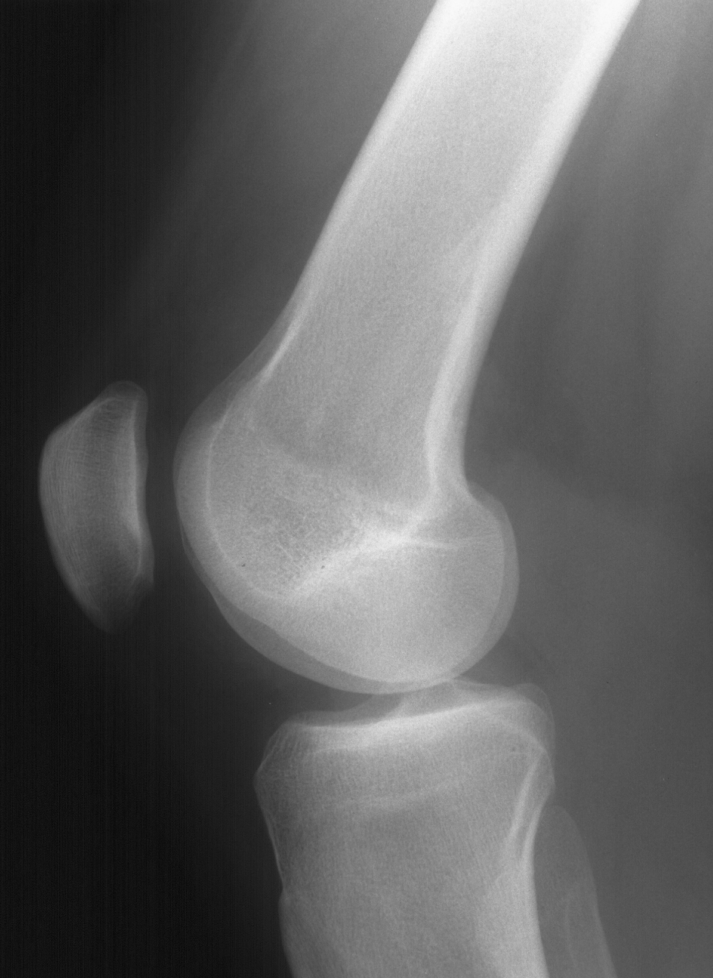
1. Lateral knee
|
1. Lateral knee

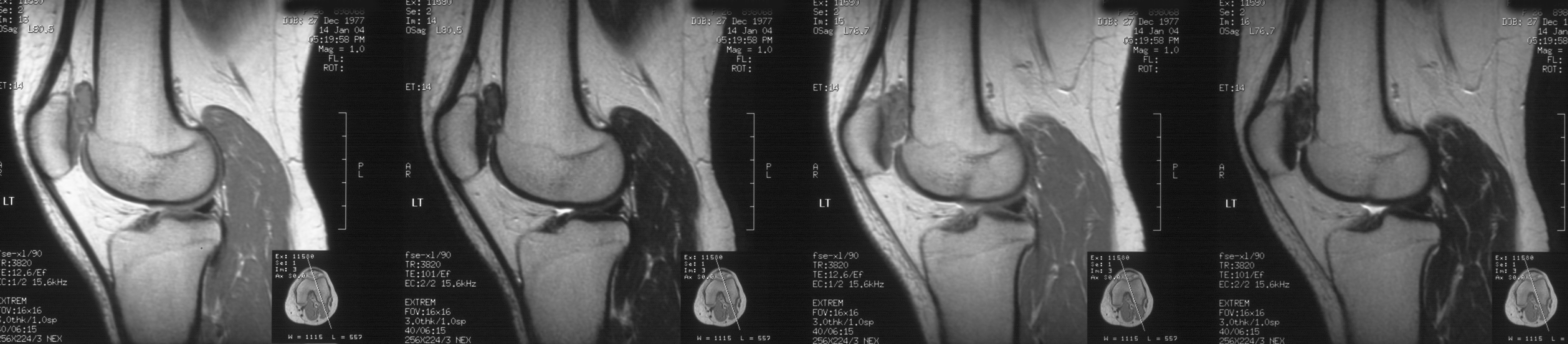
2. Sag dual echo
|
2. Sag dual echo

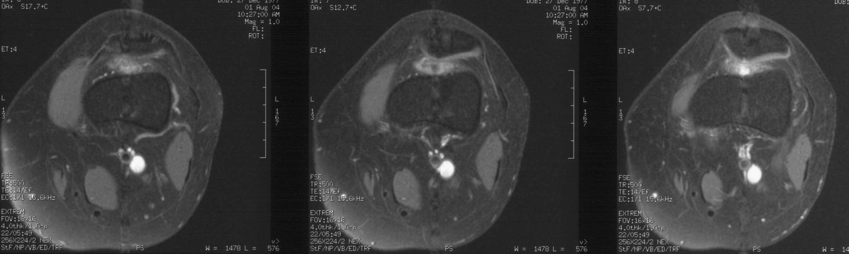
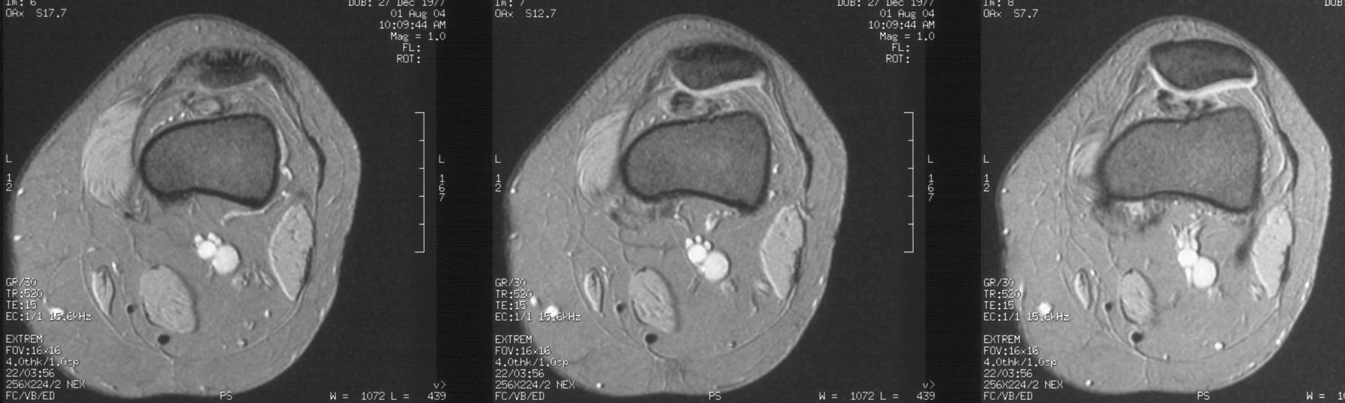
3. Ax T2W FS
|
3. Ax T2W FS